Blood Vessels Labeled Diagram - Master Blood Vessels With Quizzes And Diagrams Kenhub / More precisely, image segmentation is the process of assigning a.
Blood Vessels Labeled Diagram - Master Blood Vessels With Quizzes And Diagrams Kenhub / More precisely, image segmentation is the process of assigning a.. 2.1 brachiocephalic trunk 2.2 right common carotid 2.3 common iliac 2.4 renal. In this way, we can obtain a more. Deep veins, located in the center of the leg near the leg bones, are enclosed by muscle. An extraordinary degree of branching of blood vessels exists within the human body, which ensures that nearly every cell in the body lies within a short distance from at least one of. Blood vessels diagram this summary article displays blood vessels diagram.
Coronary circulation anatomical cross section diagram, labeled vector illustration scheme. Along with lymphatic vessels, the blood, blood vessels, and lymph, the heart composes the circulatory system of the body. Molly smith dipcnm, mbant • reviewer: Deep veins, located in the center of the leg near the leg bones, are enclosed by muscle. Blood vessels transport the blood from the heart.
The Cardiovascular System Blood Vessels And Hemodynamics Flashcards Easy Notecards from www.easynotecards.com • identification of blood vessels as arteries, capillaries or veins from the structure of their walls. 2.1 brachiocephalic trunk 2.2 right common carotid 2.3 common iliac 2.4 renal. Please click on the image(s) to view larger version. 10 photos of the the human blood vessels labeled. Label to every pixel in an image a operationalization diagram, one method of clarifing fuzzy concepts. Arteries carry oxygenated blood except in case of the pulmonary artery. Through the thin walls of the capillaries, oxygen and nutrients pass from blood into tissues. In vivo labeling of lymphendothelial cells allows parallel blood.
Blood vessels diagram | healthiack.
Blood vessels transport the blood from the heart. The blood vessel label diagram could be your consideration when creating about circulatory system. Pulmonary trunk & l/r pulm. Veins are vessels that return blood to the heart. They also take waste and carbon dioxide away from the tissues. Dimitrios mytilinaios md, phd last reviewed: The iliac, femoral, popliteal and tibial (calf) veins are the deep veins in the legs. Best quiz blood vessel labeling; Through the thin walls of the capillaries, oxygen and nutrients pass from blood into tissues. Along with lymphatic vessels, the blood, blood vessels, and lymph, the heart composes the circulatory system of the body. Automatic blood vessel segmentation in the images can help speed diagnosis and improve the diagnostic performance of less specialized physicians. All made out of smooth muscle all part of the cardiovascular system. 10 photos of the the human blood vessels labeled.
Print out these label and fill in the labels to test your blood vessels: Superior & inferior vena cava: Dimitrios mytilinaios md, phd last reviewed: Blood vessels diagram | healthiack. Exterior of the human heart.
Image Result For Human Arteries And Veins Labeled Model Anatomy Models Labeled Human Heart Anatomy Human Anatomy And Physiology from i.pinimg.com Feel free to search healthiack.com for more information on this particular topic. Dimitrios mytilinaios md, phd last reviewed: Exterior of the human heart. Circulatory system blood vessel diagram scheme on human hand silhouette. Where venules are smaller versions of veins. 960 x 720 jpeg 68 кб. Molly smith dipcnm, mbant • reviewer: We will alternatively grow the background and the vessel regions by.
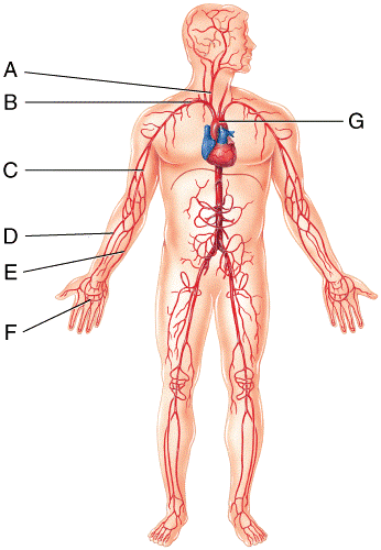
⇒ click on the diagram to show / hide labels.
⇒ click on the diagram to show / hide labels. Molly smith dipcnm, mbant • reviewer: August 17, 2020 so, you want to learn. 10 photos of the the human blood vessels labeled. In vivo labeling of lymphendothelial cells allows parallel blood. Veins are blood vessels that return blood back to the heart; To print or download this file, click the link below Deep veins, located in the center of the leg near the leg bones, are enclosed by muscle. Circulatory system blood vessel diagram scheme on human hand silhouette. (carries blood) arteries carry blood away from the heart arteries have thick and muscular walls (as they have to endure higher pressure) arteries have no valves carry oxygenated blood except for the pulmonary. October 28 observe the blood vessels diagrams above, where you can see the structures of arteries and veins clearly labeled. They also take waste and carbon dioxide away from the tissues. Capillaries are blood vessels that are one cell thick (endothelium) where the main diffusion and exchange takes place.
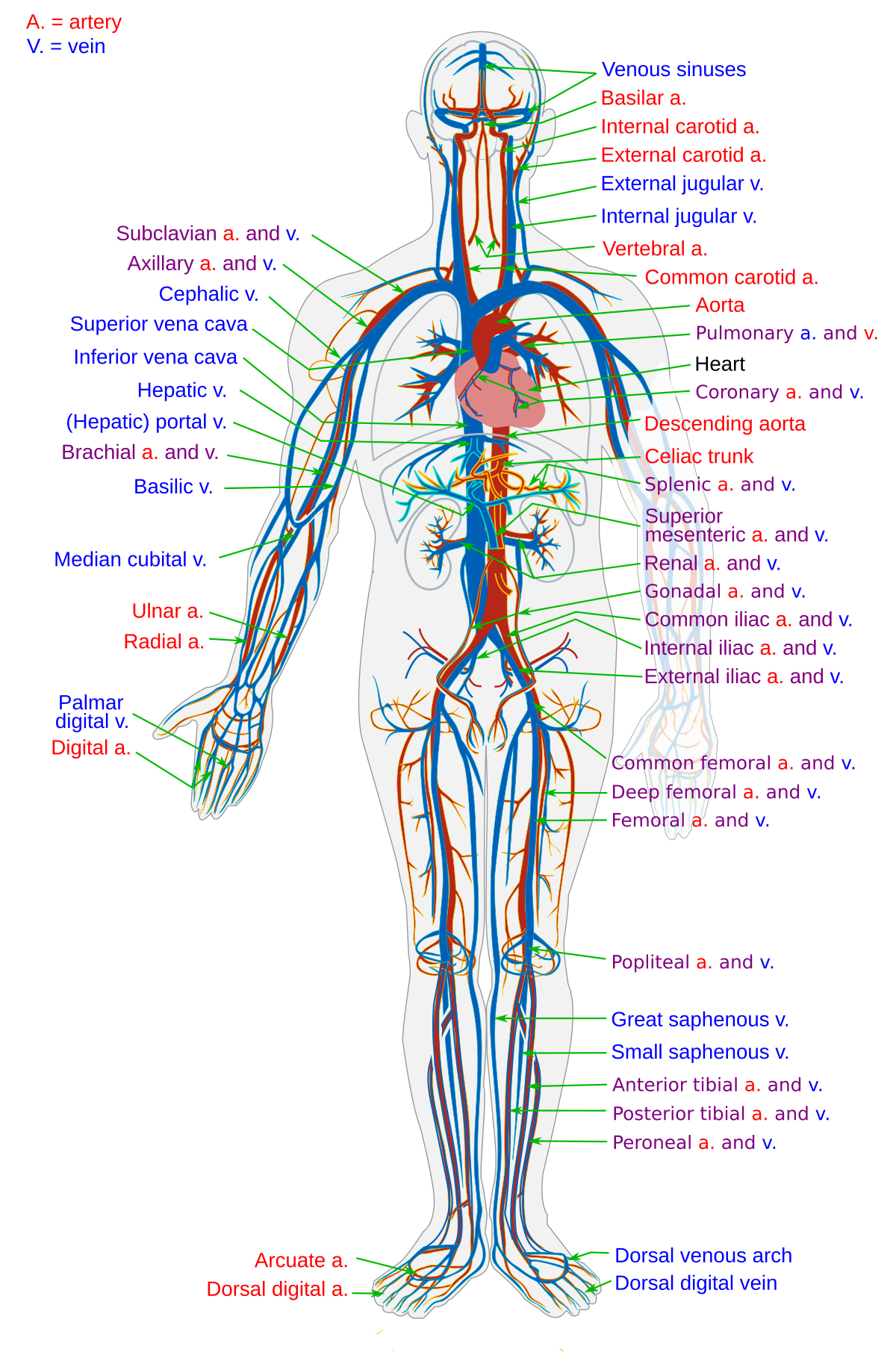
Dimitrios mytilinaios md, phd • last reviewed: Veins are blood vessels that return blood back to the heart; Automatic blood vessel segmentation in the images can help speed diagnosis and improve the diagnostic performance of less specialized physicians. Figures 1 and 2 show the major arteries and veins of the tutorials and quizzes on the circulation of blood and the anatomy, structure, and physiology of blood vessels, using interactive animations and diagrams. Arteries carry oxygenated blood except in case of the pulmonary artery.
Blood Vessel Wikipedia from upload.wikimedia.org The iliac, femoral, popliteal and tibial (calf) veins are the deep veins in the legs. The best websites voted by users. Blood vessels diagram this summary article displays blood vessels diagram. Diagram of heart with labels dissection biology, i found a number of human heart called to printable. (carries blood) arteries carry blood away from the heart arteries have thick and muscular walls (as they have to endure higher pressure) arteries have no valves carry oxygenated blood except for the pulmonary. To print or download this file, click the link below Feel free to search healthiack.com for more information on this particular topic. 1024 x 832 jpeg 293 кб.
The iliac, femoral, popliteal and tibial (calf) veins are the deep veins in the legs.
Where venules are smaller versions of veins. To print or download this file, click the link below ⇒ click on the diagram to show / hide labels. The arteries are the blood vessels which transports the blood away from the heart. Let this preset blood vessels science diagram template to show you the magic operating system of human heart. Automatic blood vessel segmentation in the images can help speed diagnosis and improve the diagnostic performance of less specialized physicians. Let's examine the anatomy of the heart along with some diagrams that show how the heart operates. Learn vocabulary, terms and more with flashcards, games and other study tools. The vessels that carry blood away from the heart are called arteries. Coronary circulation anatomical cross section diagram, labeled vector illustration scheme. Deep veins, located in the center of the leg near the leg bones, are enclosed by muscle. They also take waste and carbon dioxide away from the tissues. Learn more about the anatomy and types of blood vessels and the diseases that affect them.
In vivo labeling of lymphendothelial cells allows parallel blood blood vessels labeled. Label the following components in the diagram of the blood vessel.


0 Komentar